Osteoporosis

Osteoporosis - Vertebral fracture and kyphoplasty
The loss of bone density and the accompanying qualitative change in bone composition is called osteoporosis. It results in the fragility of bones (vertebrae, long bones) and fractures. The lifetime probability of osteoporotic fracture is 16% in women and 5% in men.
Causative factors
are considered to be smoking, taking drugs (alcohol, antiepileptics, heparin, cortisone, antiandrogens, etc.), early menopause, very low body weight and lack of physical exercise.
Every vertebral fracture in an adult is not automatically considered osteoporotic and the possibility of the presence of a bone tumour (myeloma, metastasis) with a concomitant pathological fracture should be investigated. Concomitant problems such as hyperthyroidism, hyperparathyroidism, etc. should be excluded.
Prevention of osteoporosis includes high calcium intake in childhood and adolescence and physical exercise before and after menopause, while the use of drugs and supplements is controversial in the literature.
Treatment of fractures
Treatment is initially conservative with painkillers and bed rest (7-10 days) with gradual mobilisation by a physiotherapist thereafter. In the case of severe pain pharmacoresistance, kyphoplasty has its place.
What is kyphoplasty?
It is a minimally invasive technique with a 3 mm skin incision in which a balloon is inserted and creates a cavity in the fractured vertebra. A special material (PMMA) is then injected into the vertebra , restoring its morphology and passing the pain. The procedure takes 30-45 min and the patient is mobilized the same day. Usually the elimination of pain is immediate. The same operation is also indicated in cases of vertebral fractures from myeloma or other tumours (metastatic disease). The probability of complications is considered very low 1-4%. In the following photos you can see the procedure of balloon dilation in the vertebra and the subsequent injection of the sclerosing material.
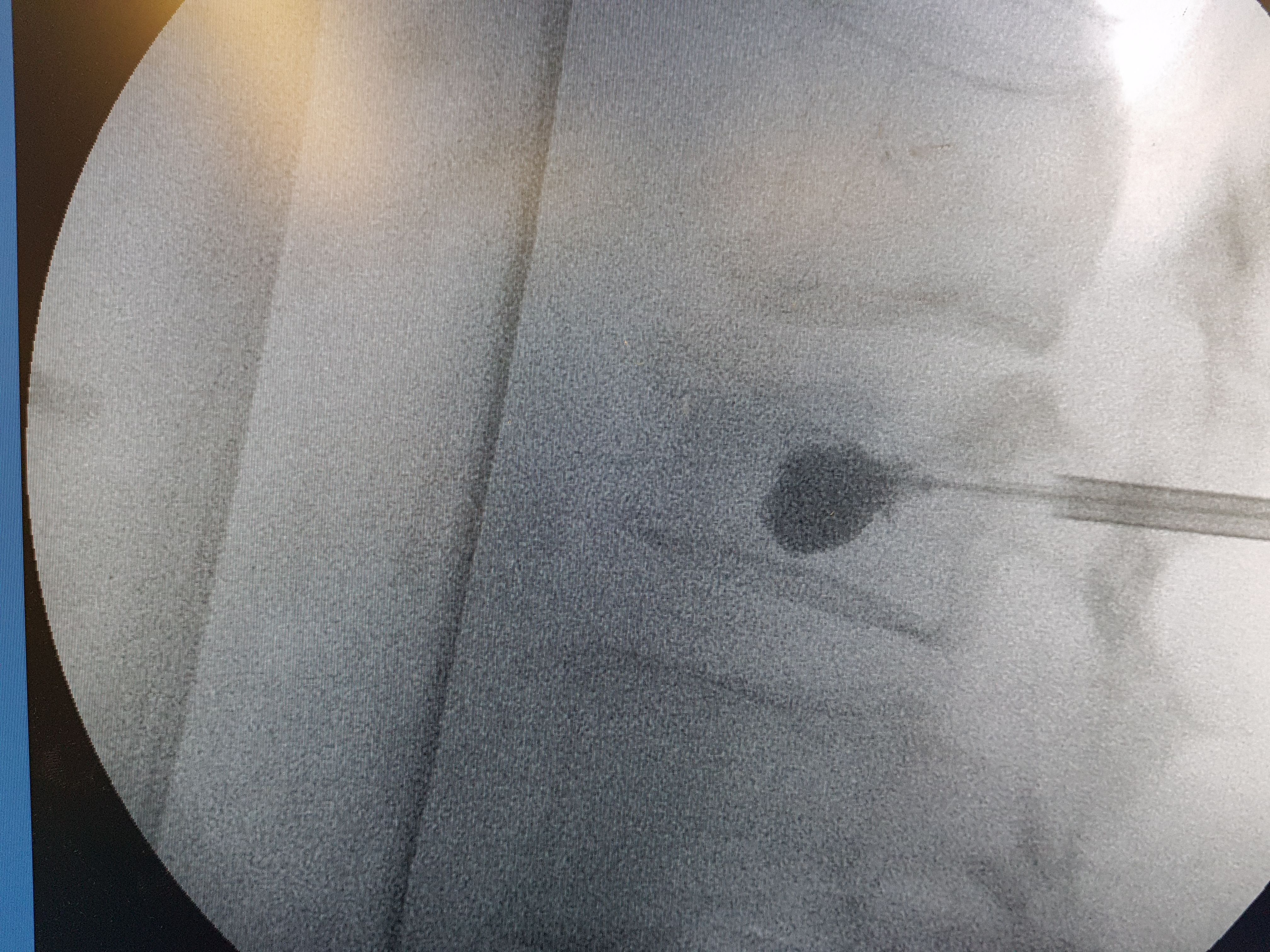 Intraoperative radiograph with the balloon extended into the vertebra.
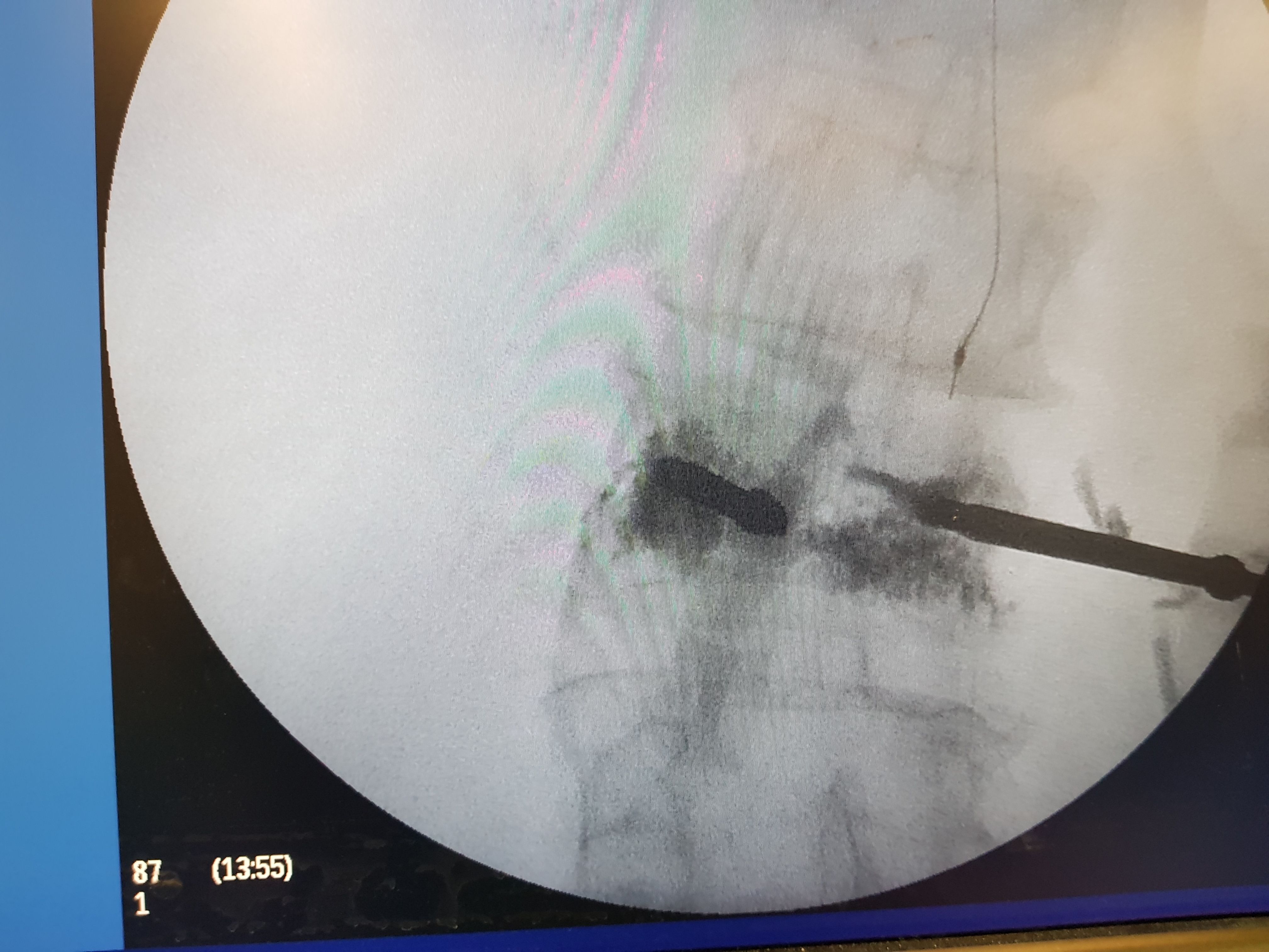 Intraoperative radiograph with the sclerosing material injected into the vertebra and restored to its shape.
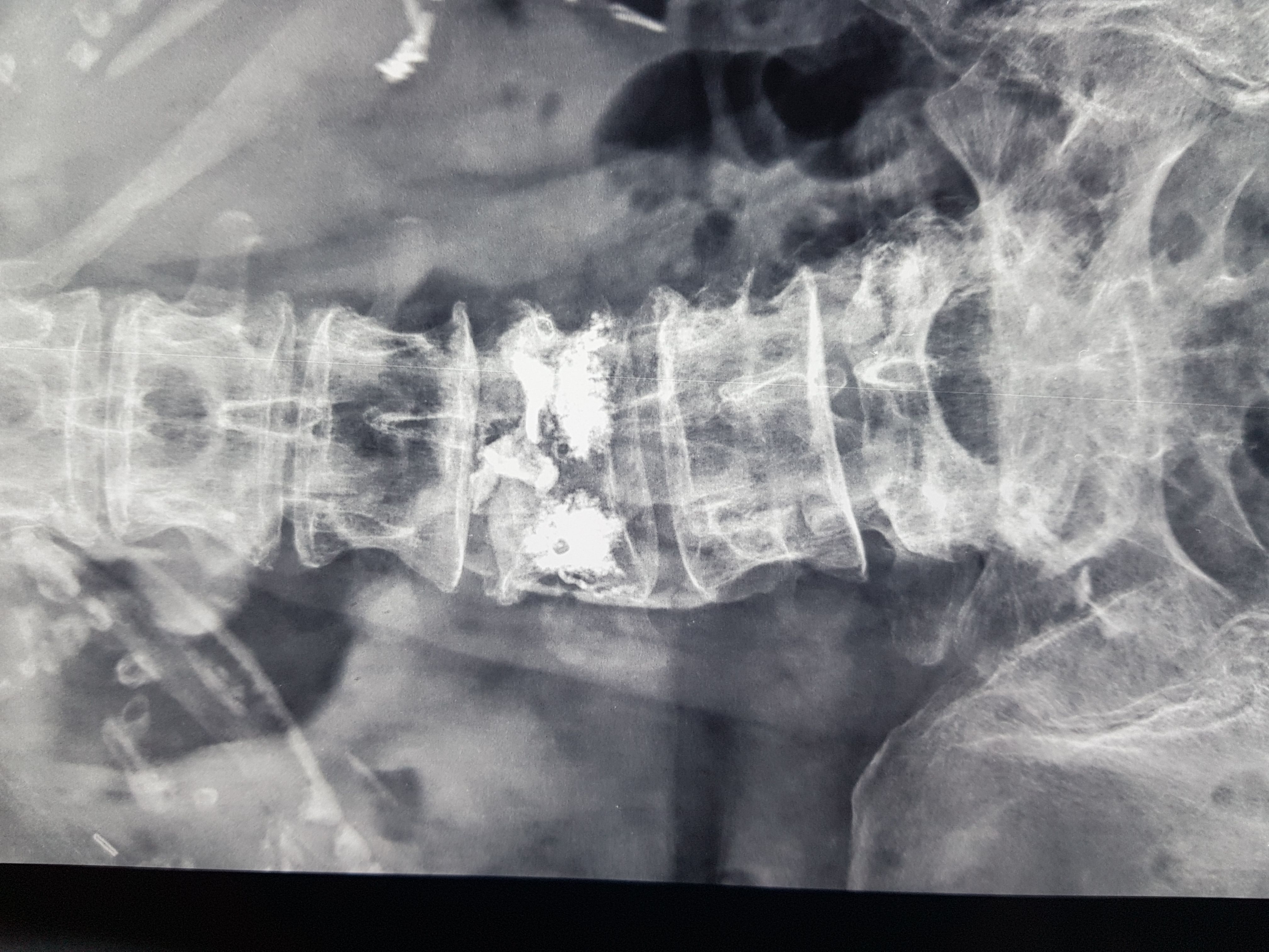 Postoperative radiograph of a patient with an osteoporotic fracture.