Brain tumours

What are brain tumours
Brain tumours are manifested by headache (55%), progressive weakness in the limbs (45%) or seizures (25%). They may be accompanied by impaired communication (dysphasia) and behavioural changes (apathy, emotional disturbance, confusion, depression, etc.)
Neurosurgical practice includes diagnosis (by stereotactic biopsy) and treatment by removal of brain tumours (gliomas, meningiomas, metastases, etc.). Surgical intervention is performed in a state-of-the-art manner using neuroplanning and brain mapping. Preoperatively in many cases we follow a special protocol that includes CT angiography scanning and embolization of the tumors so that surgical removal is possible.
Our effort is aimed at total removal of brain tumors without causing neurological complications (unfeasible in some cases). The use of modern technology (neuroplanning, mapping and intraoperative neuromonitoring) facilitates our work and the results are spectacular.
The outcome of oncological patients depends on the preoperative neurological status, type and location of the lesion, surgery, postoperative care and the oncological and radiotherapy intervention that follows.
Examples
Example 1
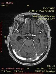
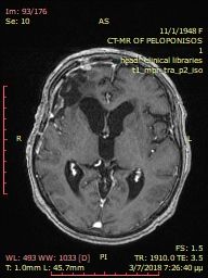
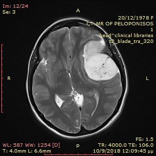
Pre-operative brain meningioma
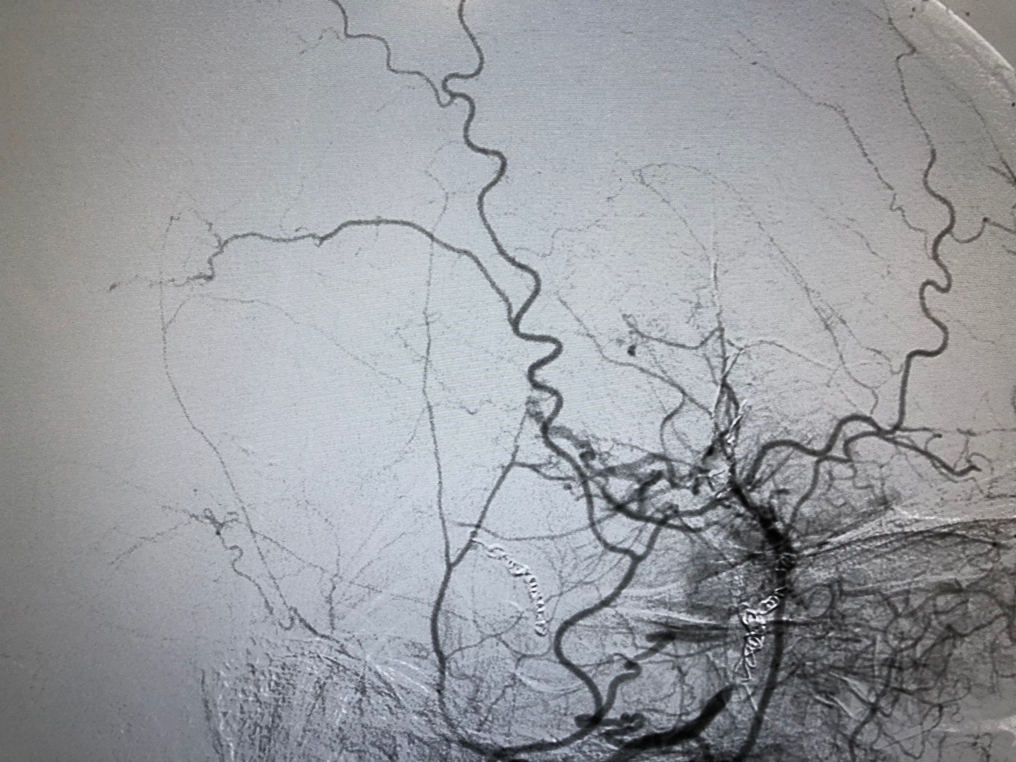
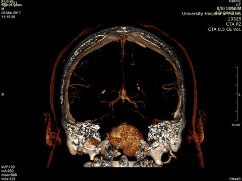
Angiography with preoperative embolization of the meningioma.
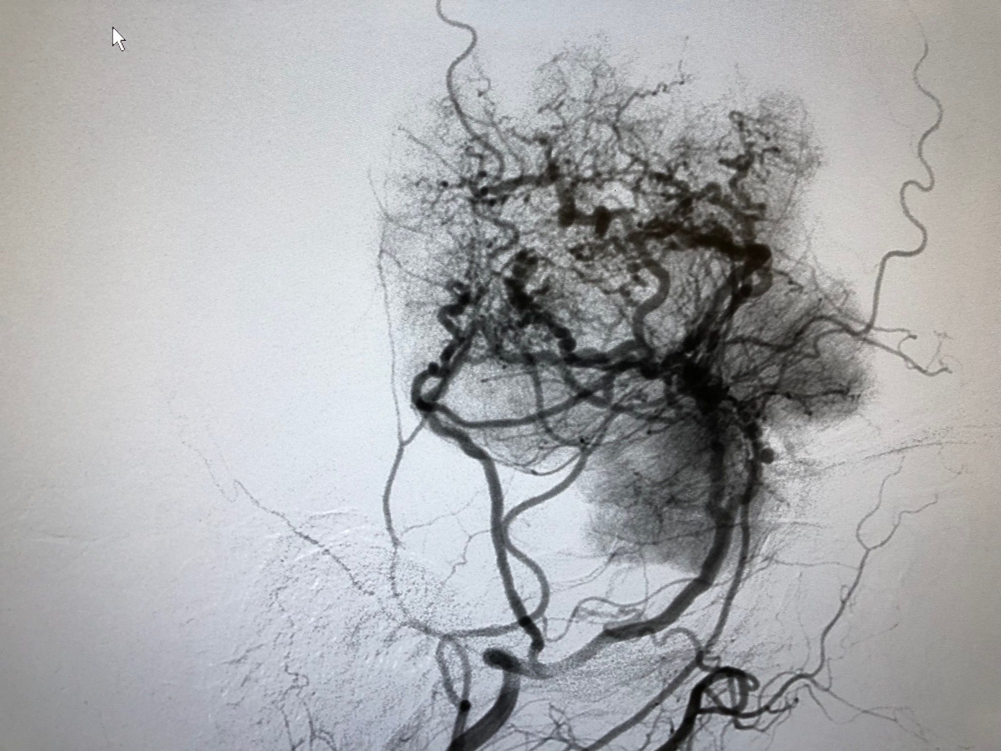
Preoperative angiography after embolization of the meningioma.
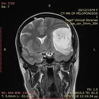
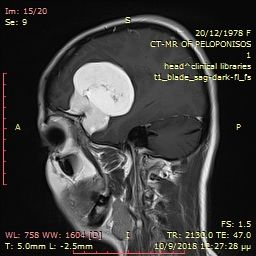
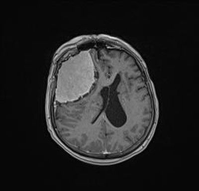
Preoperative large brain meningioma.
Pre-operative ectopic cerebral meningioma.
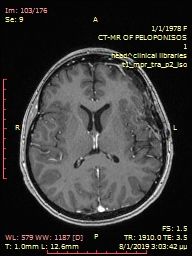
Postoperative MRI after total tumor removal.
Example 2
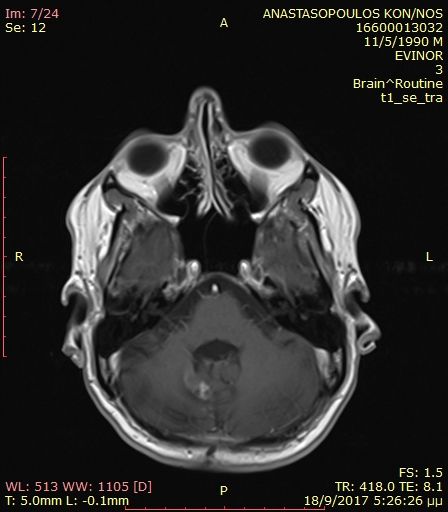
Preoperative cerebellar haemangioblastoma.
Postoperative cerebellar haemangioblastoma after total removal.
Example 3
Preoperative brain meningioma
Postoperative MRI after total tumor removal.